
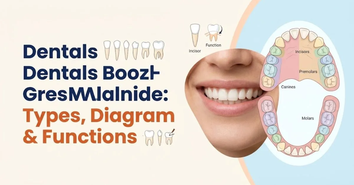
Teeth Names Complete Guide to Types, Diagram & Functions
Complete USA-focused dental anatomy guide • Tooth names, positions, numbering, functions, and oral health
Teeth names include incisors, canines, premolars, and molars, the four main types of permanent human teeth. Each tooth has a particular position, shape, and function, helping you bite, tear, crush, and grind food. Understanding these names makes dental diagrams, oral anatomy, and conversations with your dentist much easier to follow.
If you’ve ever looked at a dental chart and wondered which tooth is causing discomfort, you’re not alone. This guide explains teeth names, tooth positions, baby teeth, adult teeth, dental numbering, and tooth anatomy in straightforward language. Drawing on established dental-health references, it helps students, parents, and curious readers identify the teeth in their own mouths with greater confidence.
Human teeth are arranged in upper and lower dental arches. The four main groups are incisors, canines, premolars, and molars.
1. Types of Teeth Names
The four main types of human teeth are incisors, canines, premolars, and molars. These tooth groups differ in shape and location, but work together during chewing. Most adults have eight incisors, four canines, eight premolars, and twelve molars when all four wisdom teeth are present.
When you look in a mirror, the flat teeth at the front are incisors, while the pointed teeth beside them are canines. Premolars sit farther back, followed by the broad molars. Knowing this arrangement helps you connect common dental terms with the actual teeth you see and feel in your mouth.
Incisors
Incisors are the eight front teeth, with four in the upper jaw and four in the lower jaw. Their thin, chisel-like edges cut through food when you bite. The two middle teeth in each jaw are central incisors, while the teeth beside them are lateral incisors.
Central incisors
Central incisors are the two front teeth located directly beside the midline of the mouth in each dental arch. The upper central incisors are generally wider than the lower central incisors. They are especially important for biting into foods such as apples and sandwiches.
Lateral incisors
Lateral incisors sit immediately beside the central incisors and before the canines. There are four lateral incisors in a complete permanent dentition. Their narrower crowns help guide food toward the canines during biting, and their position contributes to the appearance of the front dental arch.
Upper and lower incisors
Upper incisors belong to the maxillary arch, while lower incisors belong to the mandibular arch. The upper and lower incisors meet during certain biting movements. Their alignment influences how the front teeth contact each other and how a person cuts food.
Canines
Canines are the four pointed teeth positioned beside the lateral incisors. Also called cuspids or eyeteeth, they help tear food and guide the bite. Each quadrant normally contains one canine, making these teeth easy to identify by their prominent shape and position between the incisors and premolars.
Why are canines called eyeteeth?
The upper canines are sometimes called eyeteeth because of their location beneath the eye region of the face. The term is a traditional anatomical nickname, not a separate tooth type. Both upper and lower canines are part of the permanent dentition and have a single prominent cusp.
Canine teeth and their roots
Canines commonly have long roots relative to many other teeth. Their roots help anchor them securely in the jaw, while the pointed crown assists with tearing food. Canines also play a role in guiding certain jaw movements, although the exact contact pattern varies between individuals.
Premolars
Premolars, sometimes called bicuspids, are located between the canines and molars. Adults normally have eight, including first and second premolars in each quadrant. Their broader chewing surfaces help crush and grind food. Unlike permanent teeth, primary or baby dentition does not contain premolars.
First premolars
First premolars are positioned directly behind the canines. Depending on the tooth, they often have two prominent cusps that help break down food. The upper first premolars and lower first premolars differ in their detailed anatomy, but both contribute to chewing.
Second premolars
Second premolars are located between the first premolars and first molars. They have broad chewing surfaces that help crush food before it reaches the larger molars. A complete permanent dentition has four second premolars, one in each quadrant.
Why premolars are called bicuspids
The term bicuspid refers to the presence of two cusps, or raised points, on many premolars. However, the exact number and arrangement of cusps vary among premolar types. Premolars are therefore identified by their location and dental classification, not simply by counting visible cusps.
Molars
Molars are the large teeth at the back of the mouth. Adults can have twelve, including first molars, second molars, and third molars. Their broad surfaces are designed for grinding food into smaller pieces. Third molars are commonly called wisdom teeth and may be absent or removed.
First molars
First permanent molars commonly erupt around age six. They emerge behind the primary teeth rather than replacing a baby tooth. Their broad chewing surfaces help grind food, and their early appearance makes them important teeth to protect during childhood.
Second molars
Second molars generally emerge farther back in the mouth during later childhood or early adolescence. They sit behind the first molars and contribute to the grinding surface of the permanent dental arch. Their eruption timing varies, so a dentist can confirm whether development is progressing normally.
Third molars or wisdom teeth
Third molars are the last molars in each quadrant and are commonly called wisdom teeth. They often emerge in the late teens or early twenties, although some people never develop all four. When there is insufficient room, a wisdom tooth may remain impacted or cause dental problems.
Quick comparison of the four teeth types
| Tooth type | Number in a complete adult set | Main function | Position |
| Incisors | 8 | Biting and cutting | Front |
| Canines | 4 | Tearing | Beside incisors |
| Premolars | 8 | Crushing and grinding | Between canines and molars |
| Molars | 12 | Grinding | Back of mouth |
| Total | 32 | Chewing and food processing | Upper and lower arches |
The total of 32 includes all four wisdom teeth. A person with missing or extracted wisdom teeth may have fewer permanent teeth.
2. How Many Teeth Do We Have? Teeth Names in Children and Adults
Most children develop 20 primary teeth, also known as baby teeth, milk teeth, or deciduous teeth. These include eight incisors, four canines, and eight molars. Adults usually have 32 permanent teeth, although the total may be lower if wisdom teeth are missing, have not erupted, or have been removed.
The difference matters because baby teeth and permanent teeth do not have identical types. Children have primary molars but no premolars. As development continues, the primary molars are replaced by premolars, while permanent molars erupt farther back in the mouth. This explains why the adult dental arch has more teeth.
Primary teeth names and numbers
| Primary tooth type | Upper jaw | Lower jaw | Total |
| Central incisors | 2 | 2 | 4 |
| Lateral incisors | 2 | 2 | 4 |
| Canines | 2 | 2 | 4 |
| First molars | 2 | 2 | 4 |
| Second molars | 2 | 2 | 4 |
| Total | 10 | 10 | 20 |
The primary dentition contains no premolars. When children lose their primary molars, permanent premolars eventually take their places.
Permanent teeth names and numbers
| Permanent tooth type | Upper jaw | Lower jaw | Total |
| Central incisors | 2 | 2 | 4 |
| Lateral incisors | 2 | 2 | 4 |
| Canines | 2 | 2 | 4 |
| First premolars | 2 | 2 | 4 |
| Second premolars | 2 | 2 | 4 |
| First molars | 2 | 2 | 4 |
| Second molars | 2 | 2 | 4 |
| Third molars | 2 | 2 | 4 |
| Total | 16 | 16 | 32 |
These are standard counts for complete primary and permanent dentitions. Individual tooth development varies, particularly for wisdom teeth.
When do baby teeth erupt?
Baby teeth typically begin emerging at around six months of age, although the timing varies considerably. The lower central incisors are commonly among the first teeth to appear. The remaining primary teeth emerge gradually as the child’s dental arches develop.
| Primary teeth | Approximate eruption period |
| Lower central incisors | 6–10 months |
| Upper central incisors | 8–12 months |
| Lateral incisors | 9–16 months |
| First molars | 13–19 months |
| Canines | 16–23 months |
| Second molars | 23–33 months |
Approximate ranges based on commonly used pediatric dental eruption charts. Individual development can differ.
By approximately three years of age, many children have a complete set of 20 primary teeth. A child’s dentist can assess eruption patterns and determine whether additional evaluation is needed.
When do permanent teeth replace baby teeth?
Permanent teeth begin appearing during early childhood, often around age six. The first permanent molars erupt behind the primary second molars, while permanent incisors gradually replace their primary counterparts.
The transition from primary to permanent teeth is called mixed dentition because both types are present in the mouth at the same time.
| Permanent tooth group | Typical development |
| First permanent molars | Around 6–7 years |
| Central incisors | Around 6–8 years |
| Lateral incisors | Around 7–9 years |
| First premolars | Around 10–12 years |
| Canines | Approximately 9–12 years, depending on arch |
| Second premolars | Around 10–12 years |
| Second molars | Around 11–13 years |
| Third molars | Commonly late teens to early adulthood |
These are general developmental ranges rather than strict deadlines. Genetics, dental development, and individual circumstances can influence eruption.
Why do children have fewer teeth than adults?
Children have fewer teeth because primary dentition is designed for the smaller jaws of early childhood. As the jaws grow, permanent teeth replace primary teeth and additional molars erupt behind them. Premolars appear as part of the permanent set, replacing primary molars rather than emerging as a separate baby-tooth group.
For parents, understanding the difference can make dental visits less confusing. A child’s first permanent molars are especially important because they erupt behind the baby teeth and are not replacements for an existing primary tooth.
3. Teeth Names and Functions
Teeth have different shapes because each group performs a different part of chewing. The front teeth cut food, the canines help tear it, and the premolars and molars break it down further. Together, the teeth, jaw muscles, and temporomandibular joints coordinate biting and chewing.
The function of a tooth is closely connected to its location. Front teeth are positioned for cutting and guiding food into the mouth, while back teeth have broader surfaces suited to grinding. When one tooth is damaged or missing, nearby teeth may be affected by changes in chewing contacts and bite alignment.
How incisors help with biting
Incisors have relatively thin crowns with sharp cutting edges. They are used when biting into foods such as fruit, sandwiches, and vegetables. Their position at the front of the mouth makes them particularly important for cutting food into smaller pieces before it is moved toward the back teeth.
The central incisors often make the first contact with food during a bite. The lateral incisors help continue the cutting process and work with the canines to guide food toward the premolars.
How canines help tear food
Canines have pointed crowns and strong roots. Their shape helps grip and tear food, while their position between incisors and premolars allows them to participate in both front-to-back transitions during chewing.
Canines also contribute to the guidance of some lateral jaw movements. In certain bite patterns, the upper and lower canines help guide the jaw as it moves sideways. The exact contact depends on the person’s dental alignment and bite.
How premolars crush and grind food
Premolars have broader crowns than incisors and canines. Their cusps and grooves create surfaces suited to crushing and grinding food. They act as a transition between the tearing action of canines and the more extensive grinding performed by molars.
For example, after biting a piece of food with the incisors, a person may move it toward the premolars. These teeth reduce its size and help prepare it for further chewing by the molars.
How molars grind food
Molars have broad chewing surfaces and multiple cusps. Their large crowns help break down food through repeated contact with opposing teeth. First and second molars generally handle much of the grinding activity in the back of the mouth.
The grooves and pits on molar surfaces can trap food particles and plaque, which is one reason careful brushing is important. Newly erupted permanent molars in children deserve particular attention because they can be difficult to reach with a toothbrush.
How teeth support speech and facial appearance
Teeth do more than process food. The front teeth help the tongue and lips form certain speech sounds, and the position of the teeth influences how air moves during speaking. Missing or misaligned front teeth may affect pronunciation for some people.
Teeth also support the appearance of the lower face and smile. Their shape, color, and alignment contribute to facial appearance, while the surrounding gums and lips frame the visible teeth.
What happens when a tooth is missing?
The effects of a missing tooth depend on its location, the surrounding teeth, and the person’s overall oral health. A missing molar can affect chewing, while a missing front tooth may affect appearance and certain speech sounds.
Adjacent teeth may shift over time, and opposing teeth can change position in some cases. A dentist can evaluate the bite, remaining teeth, and available replacement options, which may include a dental implant, bridge, or removable prosthesis when appropriate.
4. Teeth Names and Anatomy
A tooth is a specialized anatomical structure that consists of several mineralized tissues and an inner living tissue called the dental pulp. The crown is the visible portion above the gumline, while the root extends into the jaw and helps anchor the tooth.
Although teeth and bones share some mineral components, teeth are not bones. Tooth enamel is highly mineralized and protects the crown, while dentin forms much of the internal tooth structure. Cementum covers the root, and the pulp contains nerves and blood vessels.
Cross-section of a human tooth showing the crown, root, enamel, dentin, pulp, cementum, and supporting tissues.
The crown of a tooth
The crown is the portion of a tooth that is normally visible above the gumline. Its shape varies according to tooth type: incisors have cutting edges, canines have pointed cusps, and premolars and molars have broader chewing surfaces.
The crown is covered by enamel, which protects the underlying dentin. Damage to enamel can expose deeper tooth structures and may contribute to sensitivity or decay. A dentist can determine the extent of damage through an examination and, when appropriate, dental imaging.
The root of a tooth
The root anchors the tooth within the jaw. The number and shape of roots vary by tooth type. Many incisors and canines generally have one root, while molars commonly have multiple roots, although anatomical variations occur.
The root is covered by cementum and is connected to the surrounding alveolar bone through the periodontal ligament. This ligament helps support the tooth and allows slight movement under normal chewing forces.
Enamel
Enamel is the hard, highly mineralized outer covering of the tooth crown. It protects the underlying dentin against normal chewing forces and exposure to the oral environment.
Enamel does not contain living cells in the way that many other tissues do. Once significant enamel structure is lost, the body cannot naturally rebuild the original structure in the same manner as living bone tissue. Fluoride and preventive dental care can support remineralization of early mineral loss, but they do not regrow missing enamel.
Dentin
Dentin lies beneath enamel in the crown and beneath cementum in the root. It makes up much of the tooth’s structure and contains microscopic tubules that extend toward the pulp.
When enamel becomes thin or damaged, or when gums recede and root dentin becomes exposed, certain stimuli may cause sensitivity. Not every sensitive tooth has the same cause, so persistent discomfort should be assessed by a dental professional.
Cementum
Cementum is a mineralized tissue that covers the root surface. It provides an attachment surface for fibers of the periodontal ligament, which connect the tooth to the surrounding supporting tissues.
Unlike enamel, cementum is associated with living cells and can undergo limited repair or deposition. Its main role is supporting attachment rather than serving as the exposed chewing surface.
Dental pulp
The dental pulp is the soft tissue at the center of the tooth. It contains blood vessels, nerves, and connective tissue that support tooth development and sensory function.
When decay, trauma, or other damage reaches the pulp, inflammation may cause significant pain. Treatment depends on the cause and extent of the damage. A dentist may recommend a filling, pulp therapy, root canal treatment, or another appropriate procedure.
Periodontal ligament and alveolar bone
The periodontal ligament consists of connective tissue fibers that attach the tooth root to the alveolar bone. It helps support the tooth during biting and chewing and provides sensory information about forces applied to the teeth.
The alveolar bone forms the tooth sockets in the upper and lower jaws. Healthy gums, periodontal ligament, and supporting bone are essential for keeping natural teeth stable.
Are teeth bones?
No. Teeth are not bones, even though both contain mineralized tissues. Bones contain living cells and have remodeling and repair mechanisms that differ from those of teeth. Enamel, the outer tooth covering, cannot regenerate as a complete original structure after substantial loss.
The distinction is important because a chipped tooth or cavity cannot be treated like a minor bone injury. Dental damage may require restorative treatment, and the appropriate option depends on the tooth, location, and severity.
How dental anatomy differs by tooth type
| Tooth group | Typical crown characteristics | Typical root pattern |
| Incisors | Flat cutting edge | Usually one root |
| Canines | One prominent cusp | Usually one long root |
| Premolars | One or more cusps | One or two roots, depending on type |
| Molars | Broad surface with multiple cusps | Commonly multiple roots |
These are general anatomical patterns. Tooth roots, cusp arrangements, and other features vary among individuals and between upper and lower teeth.
5. What Are Teeth Numbers?
The Universal Numbering System is widely used by dentists in the United States to identify permanent teeth. It assigns each permanent tooth a number from 1 to 32, beginning with the upper-right third molar and continuing around the upper arch before moving to the lower arch.
Understanding tooth numbers can make dental appointments less confusing. When a dentist mentions a tooth number on an examination chart, that number refers to a specific tooth rather than simply its type. For example, a particular molar can be identified precisely by its number and location.
Universal Numbering System for Adult Teeth
In the US system, permanent tooth number 1 is the upper-right third molar, commonly called the wisdom tooth. Numbers continue across the upper arch to number 16, the upper-left third molar. Number 17 begins at the lower-left third molar, and the sequence ends at number 32.
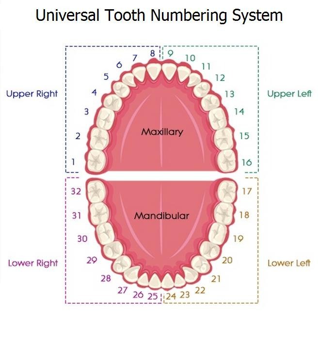
Universal Numbering System for permanent teeth, commonly used in US dental records.
Upper-right teeth: Numbers 1–8
| Number | Tooth name |
| 1 | Upper-right third molar |
| 2 | Upper-right second molar |
| 3 | Upper-right first molar |
| 4 | Upper-right second premolar |
| 5 | Upper-right first premolar |
| 6 | Upper-right canine |
| 7 | Upper-right lateral incisor |
| 8 | Upper-right central incisor |
The sequence begins at the back of the upper-right side and moves toward the center of the mouth. Tooth number 8 is the upper-right central incisor, located beside the midline.
Upper-left teeth: Numbers 9–16
| Number | Tooth name |
| 9 | Upper-left central incisor |
| 10 | Upper-left lateral incisor |
| 11 | Upper-left canine |
| 12 | Upper-left first premolar |
| 13 | Upper-left second premolar |
| 14 | Upper-left first molar |
| 15 | Upper-left second molar |
| 16 | Upper-left third molar |
The numbering continues from the upper-right central incisor across the front teeth and toward the upper-left wisdom tooth. This makes the entire upper arch a continuous sequence from 1 through 16.
Lower-left teeth: Numbers 17–24
| Number | Tooth name |
| 17 | Lower-left third molar |
| 18 | Lower-left second molar |
| 19 | Lower-left first molar |
| 20 | Lower-left second premolar |
| 21 | Lower-left first premolar |
| 22 | Lower-left canine |
| 23 | Lower-left lateral incisor |
| 24 | Lower-left central incisor |
Number 17 starts at the lower-left wisdom tooth. The sequence moves forward along the lower arch until number 24, the lower-left central incisor.
Lower-right teeth: Numbers 25–32
| Number | Tooth name |
| 25 | Lower-right central incisor |
| 26 | Lower-right lateral incisor |
| 27 | Lower-right canine |
| 28 | Lower-right first premolar |
| 29 | Lower-right second premolar |
| 30 | Lower-right first molar |
| 31 | Lower-right second molar |
| 32 | Lower-right third molar |
The final eight teeth complete the numbering system at the lower-right wisdom tooth. Together, the four groups identify all 32 possible permanent teeth.
Universal Numbering System for Baby Teeth
Primary teeth use letters rather than numbers in the US Universal Numbering System. The sequence begins with A at the upper-right second molar, moves across the upper arch to J, then continues from K at the lower-left second molar through T at the lower-right second molar.
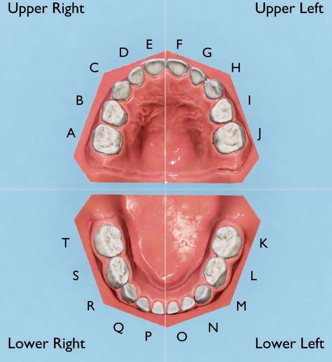
Primary teeth are identified by letters A through T in the US Universal Numbering System.
Primary upper teeth: A–J
| Letter | Tooth name |
| A | Upper-right second molar |
| B | Upper-right first molar |
| C | Upper-right canine |
| D | Upper-right lateral incisor |
| E | Upper-right central incisor |
| F | Upper-left central incisor |
| G | Upper-left lateral incisor |
| H | Upper-left canine |
| I | Upper-left first molar |
| J | Upper-left second molar |
Primary lower teeth: K–T
| Letter | Tooth name |
| K | Lower-left second molar |
| L | Lower-left first molar |
| M | Lower-left canine |
| N | Lower-left lateral incisor |
| O | Lower-left central incisor |
| P | Lower-right central incisor |
| Q | Lower-right lateral incisor |
| R | Lower-right canine |
| S | Lower-right first molar |
| T | Lower-right second molar |
The letter system helps dental professionals distinguish primary teeth from permanent teeth in patient records. If your child’s chart includes a letter, it identifies a baby tooth under this system.
How to read a dental chart
A dental chart normally shows the upper and lower dental arches, with the teeth arranged according to the patient’s right and left sides. The perspective can be confusing because a chart viewed from the front may appear reversed.
When reading your chart, first identify whether it represents the patient’s right or left. Then locate the tooth number or letter and compare it with the tooth’s position in the arch. If you are unsure, ask your dentist to point to the tooth on the chart.
What is the difference between the US and FDI numbering systems?
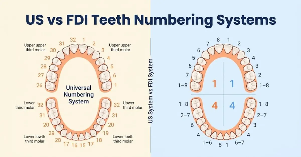
The United States commonly uses the Universal Numbering System, while many other countries use the FDI World Dental Federation system. The FDI system identifies permanent teeth using two digits: the first indicates the quadrant, and the second indicates the tooth’s position from the midline.
For example, in the FDI system, 11 identifies the upper-right central incisor, while 21 identifies the upper-left central incisor. This differs from the US Universal Numbering System, in which those teeth are numbers 8 and 9, respectively.
| Tooth | US Universal System | FDI System |
| Upper-right central incisor | 8 | 11 |
| Upper-left central incisor | 9 | 21 |
| Lower-left central incisor | 24 | 31 |
| Lower-right central incisor | 25 | 41 |
The two systems describe the same teeth using different conventions. When reviewing dental records from another country, confirm which numbering system the dentist uses.
6. Tips for Keeping Teeth Healthy
Healthy teeth depend on consistent daily care, preventive dental visits, and habits that reduce the risk of cavities and gum disease. Brushing with fluoride toothpaste, cleaning between teeth, and limiting frequent exposure to sugary foods and drinks are widely recommended oral-health practices.
Every tooth type needs attention. Incisors may be easy to reach, but the grooves of molars and the spaces between teeth can be more difficult to clean. A regular routine helps protect both the visible tooth surfaces and the areas around the gumline.
Brush twice daily with fluoride toothpaste
Brush your teeth twice a day using fluoride toothpaste and a soft-bristled toothbrush. Use gentle movements and clean the outer, inner, and chewing surfaces of the teeth. Pay particular attention to the back molars, which can be harder to reach.
Fluoride helps strengthen enamel and supports remineralization of early mineral loss. Choose toothpaste appropriate for your age and follow the product directions. For young children, caregivers should supervise brushing and use an age-appropriate amount of fluoride toothpaste.
Clean between your teeth every day
A toothbrush cannot adequately clean every surface between neighboring teeth. Floss or another suitable interdental cleaner can help remove plaque and food particles from these spaces.
Cleaning between teeth is particularly relevant around premolars and molars, where adjacent surfaces may be difficult to inspect. If conventional floss is difficult to use, a dentist or dental hygienist can recommend an alternative suited to your teeth and gums.
Limit frequent sugary snacks and drinks
Bacteria in dental plaque can use sugars to produce acids that contribute to tooth decay. Frequent exposure to sugary snacks and drinks can increase the amount of time teeth spend in an acidic environment.
Water is a practical choice between meals. If you consume sugary foods, consider the frequency of exposure and maintain your usual oral hygiene routine. Avoid using sugary beverages as a prolonged sipping habit throughout the day.
Protect newly erupted permanent molars
The first permanent molars often appear around age six, behind the primary teeth. Because they do not replace a baby tooth, parents may mistake them for primary teeth.
These molars have grooves and pits that can be difficult to clean. A dentist may recommend dental sealants for eligible children to help protect the chewing surfaces from decay.
Schedule regular dental examinations
Dental examinations allow a professional to check for cavities, gum problems, tooth eruption issues, and changes in the bite. The appropriate frequency of visits depends on individual risk factors and the dentist’s recommendations.
If you experience persistent tooth pain, swelling, sensitivity, or bleeding gums, arrange a dental evaluation. A diagram can help identify tooth names and positions, but it cannot determine the cause of symptoms or replace a clinical examination.
Protect your teeth during sports
Contact sports and activities involving falls or impacts can damage incisors, canines, and other teeth. A properly fitted mouthguard can help reduce the risk of dental injuries during many sports.
If a permanent tooth is knocked out, seek urgent dental care immediately. Handle the tooth carefully by the crown rather than the root, and follow a dentist’s or emergency dental service’s instructions while arranging treatment.
Conclusion
Knowing teeth names makes dental diagrams, oral anatomy, and conversations with a dentist easier to understand. The four main types incisors, canines, premolars, and molars work together to cut, tear, crush, and grind food. Their positions, shapes, and functions explain why each group plays a different role in everyday chewing.
A complete adult dentition normally contains 32 permanent teeth, while children typically develop 20 primary teeth. Learning tooth numbers, identifying the upper and lower arches, and understanding the difference between baby and permanent teeth can help you read dental charts with greater confidence.
Whether you’re a student learning dental anatomy, a parent following your child’s tooth development, or someone preparing for a dental appointment, this teeth names guide provides a foundation for identifying the teeth and understanding their functions. For individual dental concerns, consult a qualified dentist who can examine your teeth and recommend appropriate care.
FAQs
These frequently asked questions cover common search queries about human teeth names, their positions, and their functions.
What are the 32 teeth names?
The 32 permanent teeth consist of eight incisors, four canines, eight premolars, and twelve molars. The molars include four first molars, four second molars, and up to four third molars, commonly called wisdom teeth.
What are the 4 front teeth called?
The four front teeth in each dental arch are two central incisors and two lateral incisors. The upper arch has four incisors, and the lower arch has four, making eight permanent incisors altogether.
What are the 8 front teeth called?
The eight permanent front teeth are four central incisors and four lateral incisors. They are located in the upper and lower jaws and help cut food during biting.
What are the teeth next to the front teeth called?
The teeth immediately beside the lateral incisors are the canines. There are four permanent canines, one in each quadrant. They have pointed crowns that help tear food.
What are the back teeth called?
The back teeth include premolars and molars. Premolars are positioned behind the canines, while molars sit farther back. Wisdom teeth are the third molars at the back of the permanent dental arch.
What are the teeth beside your canine teeth called?
The teeth immediately behind the canines are the first premolars. The second premolars are located behind the first premolars and before the first molars.
What are the names of the upper teeth?
The upper teeth are part of the maxillary dental arch. In each half of the upper arch, a complete permanent set includes a central incisor, lateral incisor, canine, first premolar, second premolar, first molar, second molar, and third molar.
What are the names of the lower teeth?
The lower teeth are part of the mandibular dental arch. Each half normally contains a central incisor, lateral incisor, canine, two premolars, and three molars when all permanent teeth are present.
What are baby teeth called?
Baby teeth are called primary teeth, deciduous teeth, or milk teeth. A complete primary dentition contains 20 teeth: eight incisors, four canines, and eight molars.
Which teeth are called wisdom teeth?
Wisdom teeth are the third molars. Most people can develop up to four wisdom teeth, one in each quadrant, although some people develop fewer or none.
Are premolars and bicuspids the same?
Yes. Premolars are commonly called bicuspids because many have two prominent cusps. They are permanent teeth located between the canines and molars and help crush and grind food.
What is the difference between a tooth crown and a root?
The crown is the portion of the tooth normally visible above the gumline. The root extends into the jaw and anchors the tooth. Enamel covers the crown, while cementum covers the root.
Which teeth are numbered 1 and 32?
In the US Universal Numbering System, tooth number 1 is the upper-right third molar, and tooth number 32 is the lower-right third molar. Both are wisdom teeth.
Do all adults have 32 teeth?
No. Thirty-two is the number in a complete permanent dentition that includes four wisdom teeth. Some adults have fewer teeth because wisdom teeth are absent, have not erupted, or have been removed. Other teeth may also be missing or extracted.

Liam Anderson is a passionate cat lover and the creator of OringeCatsNames. He enjoys sharing unique orange cat name ideas, pet tips, and helpful content to make life easier for cat owners around the world.






One Comment